What Is the OCULUS Pentacam?
The OCULUS Pentacam is the industry gold standard in anterior segment tomography — a rotating Scheimpflug camera system that captures a comprehensive 3D map of the entire anterior eye segment in under two seconds. Developed by OCULUS Optikgeräte GmbH (Wetzlar, Germany), the Pentacam family has been the reference instrument for corneal analysis in refractive, cataract, and contact lens practices for over 20 years, with its software continuously validated through thousands of peer-reviewed clinical studies.
Software version 1.25r15 represents the mature, stable release of the Pentacam software platform, incorporating the full suite of diagnostic displays including the Belin/Ambrósio Enhanced Ectasia Display, Belin ABCD Keratoconus Staging and Progression, contact lens fitting, corneal densitometry, wavefront analysis, and IOL calculation modules.
Core principle of Pentacam imaging: Unlike Placido-based topographers that analyze only reflected rings from the tear film surface, the Pentacam uses Scheimpflug photography to capture true cross-sectional images of the entire anterior segment — anterior and posterior corneal surfaces, crystalline lens, and anterior chamber — independent of tear film quality. This makes it the only technology that directly measures the posterior corneal surface, which is critical for accurate ectasia detection and true corneal power calculation.
The Pentacam Hardware Family
The Pentacam software runs on several hardware configurations:
| Model | Key Differentiation |
|---|---|
| Pentacam | Standard model — 25,000 measuring points, rotating Scheimpflug system |
| Pentacam HR | High-Resolution — 138,000 measuring points, 1.45 MP camera, 100 Scheimpflug images per scan, 5× resolution of standard |
| Pentacam AXL | HR + integrated axial length measurement for one-stop IOL calculation |
| Pentacam AXL Wave | AXL + total wavefront + refraction + retroillumination |
| Pentacam Cornea OCT | HR + Spectral Domain OCT for epithelial mapping |
The Pentacam HR is the reference model for demanding refractive and cataract surgery practices. The software modules described below apply across the family, with some features exclusive to HR and newer generations.
How Scheimpflug Tomography Works
The Pentacam rotates 360° around the patient’s optical axis while capturing slit-lamp Scheimpflug images. Key advantages over competing technologies:
Tear-film independence: Measurements capture actual tissue — not reflections from the tear film surface. A patient with dry eye, irregular tear film, or surface pathology can be measured accurately.
Posterior corneal measurement: The Pentacam directly measures the posterior corneal surface — impossible with Placido topographers. This is essential for detecting early posterior ectasia (the hallmark of subclinical keratoconus), calculating true corneal refractive power (TCRP), and selecting premium toric IOLs.
Full anterior segment capture: A single scan simultaneously captures corneal topography (anterior and posterior), pachymetry, anterior chamber depth and volume, chamber angle, crystalline lens opacity staging, and iris imaging — all in under 2 seconds.
Measurement quality: Automatic quality assessment flags scans with poor fixation, blink artifacts, or inadequate illumination. The system will not display unreliable data.
Basic Software Package — Included in All Systems
General Overview Display
The primary clinical dashboard for rapid patient assessment:
- Keratometry values (K1, K2, Km, Kmax) — simulated and Scheimpflug-based
- Pachymetry: minimum corneal thickness point with location, apex thickness, central thickness
- Anterior chamber depth (ACD), volume (ACV), and angle (ACA)
- Integrated glaucoma risk assessment: three parameters (ACD, ACV, ACA) displayed together
- Scheimpflug image of the full anterior segment
- Quality Score for measurement reliability
Fast Screening Report (Indices Report)
Designed for efficient clinical workflow — all critical indices at a glance:
- Automated summary of abnormalities detected during the scan
- Directs clinician attention to areas of concern
- Risk indicators for keratoconus, ectasia risk, and asymmetry
- Suitable for delegation to trained technicians for preliminary screening
Scheimpflug Image Overview
- Full-resolution cross-sectional images of the anterior segment
- Visualization of corneal opacities, anterior segment pathology, IOL position
- Intuitive patient communication tool — patients can see their own eye structure
- Detects narrow angles, early cataracts, and implant positioning
Virtual Eye
- Rotatable, pivotable 3D model of the anterior eye segment
- Built from the patient’s actual measurement data
- Invaluable for patient education and consent discussions
- Allows visualization of corneal abnormalities from any angle
Tomography Display
- Full tomographic representation of corneal structure
- Elevation maps of anterior and posterior surfaces
- Pachymetry maps (absolute and relative)
- True anterior segment cross-sections
Iris Image
- High-resolution iris photograph with keratometry overlay
- Toric IOL alignment axis marked directly on iris image
- Critical for planning toric IOL orientation and axis verification during surgery
Topometric / 4 Maps Refractive Display
- Combined corneal curvature and power map display
- Sagittal and tangential curvature of both corneal surfaces
- Refractive power maps for clinical assessment and surgical planning
- Standard 4-map format familiar to refractive surgeons
Keratoconus and Ectasia Detection
The Pentacam’s ectasia detection capabilities are its most clinically validated feature, with decades of published clinical evidence.
Belin/Ambrósio Enhanced Ectasia Display (BAD)
Developed by Prof. Michael Belin and Prof. Renato Ambrósio Jr., the BAD display is considered the most sensitive single screening tool for subclinical keratoconus and post-LASIK ectasia risk:
- Enhanced Reference Surface: Compares the measured corneal shape to a best-fit reference surface — deviations from this surface indicate focal ectasia even before curvature changes become visible
- Multi-parameter analysis: Integrates anterior surface elevation, posterior surface elevation, pachymetry distribution, and thickness progression into a single composite index (D-value)
- D-value: The composite ectasia index — deviations >1.6 indicate high suspicion for ectasia; values between 1.0 and 1.6 require careful consideration
- Color coding: Each parameter is displayed with Z-scores relative to a normal population, with red indicating values outside normal limits
Belin ABCD Keratoconus Staging and Progression Display
The modern replacement for the Amsler-Krumeich classification, approved by the four major international ophthalmology societies:
- A — Anterior radius of curvature (3mm zone)
- B — Back (posterior) radius of curvature (3mm zone)
- C — Corneal thickness at the thinnest point
- D — Distance Corrected Visual Acuity (DCVA)
The ABCD system detects progression from 4 independent parameters — including posterior curvature changes that precede anterior surface changes in early keratoconus. Up to 8 exams per eye can be overlaid to track progression longitudinally.
Belin ABCD Progression Display: Graphs all 4 parameters over time with 80% and 95% confidence intervals. After corneal crosslinking (CXL), the display shows a separation point allowing objective documentation of treatment response.
Topometric / KC Staging Display
- Fourier analysis of the anterior corneal surface
- Spherical, astigmatic, and irregular components quantified separately
- Decentration analysis: asymmetry index >0.43 raises suspicion for keratoconus
- Automatic classification against normal population data
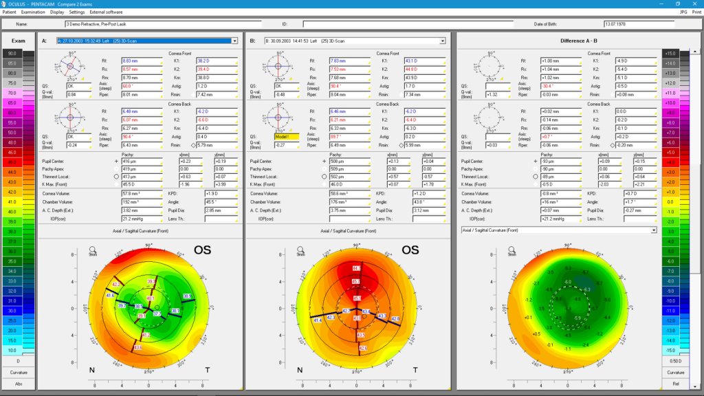
Comparison Displays
- Up to 4 examinations loaded simultaneously for direct progression monitoring
- Map subtraction to visualize changes between examinations
- Before/after CXL comparison with objective documentation
- Pre/post-LASIK surface analysis
IOL Calculation and Cataract Planning
Cataract Pre-Op Display
The comprehensive pre-operative planning dashboard for premium IOL selection:
- True Corneal Refractive Power (TCRP) calculated via ray tracing — accounts for anterior AND posterior corneal surfaces
- Assessment of corneal power with adjustable calculation zones
- 4-step evaluation of corneal optical quality for premium IOL selection
- Full anterior chamber characterization for IOL sizing
- Scheimpflug-based Pentacam Nucleus Staging (PNS) for cataract density quantification
Holladay Report
Developed in collaboration with Prof. Jack T. Holladay (USA):
- Specialized for IOL power calculation after prior corneal refractive surgery (LASIK, PRK, RK)
- Critical when pre-operative data is unavailable — uses posterior corneal data to infer pre-surgical K values
- Equivalent Keratometry Readings (EKRs) in multiple zones
- Calculates true corneal power using the relationship between anterior and posterior surfaces
IOL Calculator Module (Optional)
- Over 350 IOL geometries from major manufacturers
- Multiple power calculation formulas including advanced formulas
- Automatic transfer of K measurements — eliminates transcription errors
- Links to external calculators: PhacoOptics, OKULIX, BESSt II formula for challenging post-refractive cases
- Toric IOL planning with iris image keratometry overlay for axis marking
Phakic IOL (pIOL) Simulation and Planning
For iris-fixated phakic IOL implantation:
- Pre-operative anterior chamber evaluation for patient selection
- Calculation of required pIOL refractive power using the van der Heyde formula
- pIOL simulation including aging prediction — visualizes future crystalline lens growth and IOL clearance
- Critical for ensuring adequate endothelial clearance over the patient’s lifetime
Contact Lens Fitting Software
Standard Contact Lens Fitting
- Fluorescein pattern simulation for rigid gas-permeable (RGP) lens fitting
- Dynamic simulation of lens seat and inclination
- Prediction of lens behavior before physical fitting
Scleral Lens Fitting
- Cornea Scleral Profile (CSP) measurement up to 22mm — covers cornea AND sclera
- Sagittal height measurements for scleral lens sizing
- Tear layer clearance mapping for scleral lens optimization
- Coverage for all ages and highly abnormal corneas (keratoconus, post-keratoplasty)
Orthokeratology Monitoring
- Precise assessment of corneal changes induced by ortho-k lenses
- Follow-up comparison displays for treatment monitoring
- Epithelial redistribution tracking
Corneal Densitometry
Objective, quantitative analysis of corneal transparency:
- Color-coded opacity map across full corneal depth and area
- Assessable in different layers and zones (anterior, central, posterior stroma; epithelium)
- Applications:
- Fuchs endothelial dystrophy monitoring
- Corneal scar quantification
- Pre/post-CXL density changes
- Corneal haze after PRK or LASIK
- Contact lens-induced hypoxia
- Objective documentation for treatment planning and insurance reports
Wavefront Analysis and Aberrometry
Corneal Wavefront (Zernike Analysis)
- Elevation-based Zernike analysis of the anterior corneal surface
- Ray-traced wavefront for anterior, posterior, and total cornea
- Higher-order aberrations calculated and displayed with normative comparison
- Z-score deviation from normal population for immediate clinical interpretation
- Not possible with Placido-based topographers (which cannot measure posterior surface contribution)
Power Distribution
- Visual representation of corneal refractive power considering both surfaces
- Displays the actual corneal power distribution across the pupil
- Critical for premium multifocal and extended-depth-of-focus IOL planning
DICOM Integration and Workflow
- Full DICOM compatibility — Pentacam software integrates with hospital and clinic PACS systems
- DICOM Modality Worklist (MWL) — automatically imports patient data from the HIS/EMR
- Eliminates manual patient data entry and associated errors
- Direct data transfer to EMR/EHR systems
- Compatible with major ophthalmic practice management software platforms
Clinical Applications by Subspecialty
Refractive Surgery (LASIK, PRK, SMILE)
Pre-operative: ectasia screening (BAD display), corneal thickness assessment (sufficient residual bed), anterior chamber evaluation. Post-operative: monitoring for regression, haze, or induced ectasia.
Cataract Surgery
Premium IOL planning (toric, multifocal, EDOF) using TCRP and iris imaging. Challenging post-refractive cases using the Holladay Report. Cataract staging with PNS for surgical planning.
Keratoconus Management
Detection, staging (ABCD), and progression monitoring. CXL candidate selection and treatment response documentation. Fellow eye surveillance in unilateral cases.
Contact Lens Practice
All modalities: RGP, scleral, orthokeratology. Particularly valuable for keratoconus patients requiring specialty lens fitting.
Glaucoma Screening
Anterior chamber angle, depth, and volume as risk factors for angle-closure glaucoma. Corneal thickness (CCT) for IOP correction and glaucoma risk stratification.
Corneal Disease Monitoring
Fuchs dystrophy, corneal scars, corneal edema, post-keratoplasty. Objective densitometry for quantitative progression documentation.
Pentacam Software Modules Overview
| Module | Basic | Optional | Application |
|---|---|---|---|
| General Overview | ✅ | — | Daily screening |
| Fast Screening Report | ✅ | — | Quick triage |
| Scheimpflug Image Overview | ✅ | — | Pathology visualization |
| 4 Maps Refractive | ✅ | — | Curvature analysis |
| Virtual Eye (3D model) | ✅ | — | Patient education |
| Tomography | ✅ | — | Structural analysis |
| Topometric / KC Staging | ✅ | — | Keratoconus detection |
| Belin/Ambrósio BAD Display | ✅ | — | Ectasia screening |
| Belin ABCD Progression | ✅ | — | KC monitoring |
| Corneal Densitometry | ✅ | — | Opacity analysis |
| Comparison Displays | ✅ | — | Progression tracking |
| Cataract Pre-Op Display | Optional | ✅ | Premium IOL planning |
| Holladay Report | Optional | ✅ | Post-refractive IOL |
| IOL Calculator | Optional | ✅ | Intraocular lens power |
| pIOL Simulation | Optional | ✅ | Phakic IOL planning |
| Contact Lens Fitting | Optional | ✅ | CL practice |
| Scleral Lens Module | Optional | ✅ | Scleral lens fitting |
| Corneal Wavefront | Optional | ✅ | Aberrometry |
| Power Distribution | Optional | ✅ | Premium IOL selection |
Frequently Asked Questions
What is the clinical difference between Pentacam tomography and Placido disc topography? Placido topography measures only the anterior corneal surface by analyzing reflections of concentric rings from the tear film. Results depend on tear film stability and cannot capture the posterior corneal surface. Pentacam Scheimpflug tomography captures direct cross-sectional images of all tissue layers — anterior cornea, posterior cornea, stroma, and anterior chamber — independent of tear film. Only tomography can detect posterior ectasia (the earliest sign of keratoconus), accurately calculate true corneal power including posterior contribution, and measure anterior chamber parameters.
Can the Pentacam detect keratoconus before it is visible on curvature maps? Yes. The Belin/Ambrósio Enhanced Ectasia Display (BAD) and the ABCD Staging system analyze posterior corneal surface changes and corneal thickness distribution patterns that precede anterior curvature changes. Subclinical keratoconus — affecting an eye that appears normal on curvature maps — can often be detected through posterior elevation and thickness progression patterns.
Is the Pentacam measurement affected by contact lens wear? Yes. Rigid gas-permeable lens wear temporarily induces corneal warpage that alters topographic measurements. Patients should discontinue rigid lenses (minimum 1 week for RGP, 3 weeks for scleral) before pre-operative evaluation. Soft lenses require 3 days minimum. The Pentacam’s tear-film independence is not affected by contact lens history; it relates only to acute tear film surface irregularity at the time of measurement.
How accurate is the Pentacam for IOL power calculation after LASIK? The Pentacam’s ability to measure the posterior corneal surface directly makes it superior to standard keratometry for post-LASIK IOL calculation. The Holladay Report uses this posterior surface data to calculate effective corneal power when pre-operative data is unavailable. External calculators (BESSt II, Phaco Optics) can receive Pentacam data via direct links.
How many comparison exams can be loaded simultaneously? Up to 4 examinations can be loaded and analyzed in most comparison displays for progression assessment. The Belin ABCD Progression Display can show up to 8 examinations per eye, making it the most comprehensive keratoconus progression tracking tool available.
Summary
The OCULUS Pentacam software platform — including version 1.25r15 — is the most comprehensively validated anterior segment tomography software in clinical ophthalmology, backed by two decades of peer-reviewed research and adopted as the reference standard by leading refractive and cataract surgeons worldwide.
Its combination of tear-film-independent Scheimpflug imaging, direct posterior corneal measurement, the clinically validated Belin/Ambrósio Enhanced Ectasia Display, Belin ABCD Keratoconus Staging and Progression tracking, true corneal refractive power calculation, and integration with IOL calculators and contact lens fitting tools makes it the single most information-dense diagnostic instrument in the modern refractive surgery practice.
Get a license — free consultation
Pricing depends on version and number of users. Message us on Telegram and we’ll reply with an exact quote — no commitment required.
|
✓
20+ years experience
Software engineers with a long track record
|
⚡
Delivered within 24h
Your license is sent within one business day
|
↩
Money-back guarantee
If the license doesn’t work, we refund in full
|
Usually reply within a few hours — free consultation, no upfront payment